


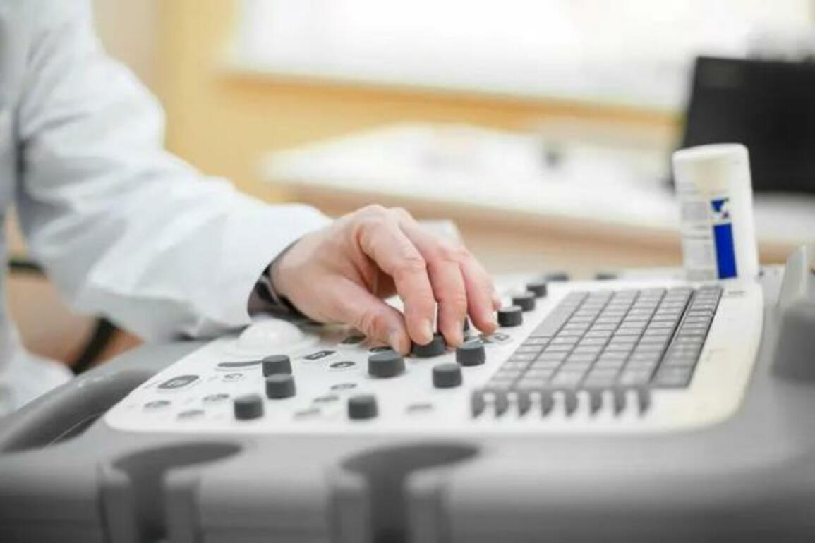
Echocardiography method and its benefits
Echocardiography or Cardiac Ultrasound uses ultrasound waves to visualize the heart, heart valves, and major vessels. Echocardiography can help clarify the thickness of the heart walls (hypertrophy or atrophy), assess their motion, and suggest the presence of ischemia or MI.
Echocardiography can assess both systolic and diastolic filling capacity of the left ventricle. Also, it may assist in evaluating left ventricular hypertrophy, hypertrophic or restrictive cardiomyopathy, severe heart failure, and constrictive pericarditis. It is also used to evaluate valve structure and function; detect valve vegetations and intracardiac thrombi, and assess pulmonary artery pressure and central venous pressure.
Echocardiography Techniques
There are different techniques for performing echocardiography:
Transthoracic echocardiography (TTE) is the most common echocardiography technique. The sensor is placed in multiple areas on the front chest wall around the heart. TTE is the most common technique, allowing to obtain two-dimensional images of large cardiac structures. Transthoracic echocardiography is a relatively inexpensive and noninvasive imaging technique for diagnosing right and left ventricular function abnormalities, movement of their walls, size and anatomy of cardiac cavities, valve system functioning, aortic root structure, and intracardiac pressure.
Transesophageal echocardiography (TEE)
The probe is placed at the end of an endoscope, allowing the heart through the stomach and esophagus to be viewed and studied.
Transesophageal echocardiography is used to visualize cardiac structures when TTE is technically tricky, such as in patients with obesity and chronic obstructive pulmonary disease (COPD). Small abnormal structures (e.g., vegetations in endocarditis or open oval window) and posterior structures (e.g., left atrium, left atrial auricle, interatrial septum, pulmonary vein anatomy) are better visualized with TEE because they are closer to the esophagus than to the front chest wall. Transesophageal echocardiography can also be used to image the ascending aorta, small masses (such as thrombi, vegetations), and artificial valves.
Stress Echocardiography
Stress Echocardiography is an alternative to radionuclide imaging for detecting myocardial ischemia during and after exercise or pharmacological stress testing. This technique detects regional abnormalities of cardiac wall motion resulting from abnormal blood flow in the epicardial vessels of the heart during the exercise test.
Computer programs estimate step-by-step ventricular contraction during systole and diastole, during rest and exercise or load. The protocols for the stress studies are similar to those for the radionuclide stress test, with the difference that dobutamine is the preferred pharmacological agent over dipyridamole.
Stress echocardiography is an important method for investigating the severity of the hemodynamic impairment in aortic valve stenosis in symptomatic patients with a low resting transvalvular pressure gradient.
Echocardiography - Indications for use
Echocardiography (Cardiac Ultrasound) diagnoses various conditions in a patient. The reason for the procedure may be the following symptoms in a person:
- Heart Murmurs, Rhythm Disturbances;
- Signs Indicating The Development Of Heart Failure, Such As Swelling Of The Extremities, Pain In The Liver;
- Acute Or Chronic Myocardial Infarction;
- Chronic Fatigue, Shortness Of Breath;
- Frequent Colds Or Fever Without Signs Of Acute Respiratory Infections;
- Predisposition To Cardiovascular Disease;
- Fainting And Angina Attacks and many more.
In addition, the indications include Rheumatic diseases, high blood pressure, and conditions accompanied by pain and numbness in the left arm, scapula, and forearm. The method is also used to monitor the effectiveness of treatment for various heart diseases and also before the upcoming surgical intervention. For prevention, it is recommended to proceed with a cardiac ultrasound examination of persons whose working activity is related to frequent emotional or physical strain.
Application among pregnant women
During pregnancy, women are prone to many diseases. Due to the changes occurring in the body, the load on the heart also increases.
Are there any contraindications?
Echocardiography has no absolute restrictions, but some recommendations should always be followed during the diagnosis.
- An echocardiogram should be performed 2-3 hours after a meal. When the stomach is full, the diaphragm may put pressure on the heart, which will affect the accuracy of the data obtained;
- It is recommended to postpone the procedure for those patients who have open wounds or severe skin diseases in the chest area;
- If the chest is deformed, the diagnostic results may be inaccurate.
- Transesophageal echocardiography (TEE) should not be used in patients with an increased gag reflex, psychiatric disorders, or esophageal abnormalities.
Athens cardiologist Dr. Ilham Kaffa
kaffailham.gr
DIONISIOS AREOPAYTOU 3
TK 11742, ATHENS
Area Makrygianni - Koukaki
Tel.: 210 9210423 Click here for further information